
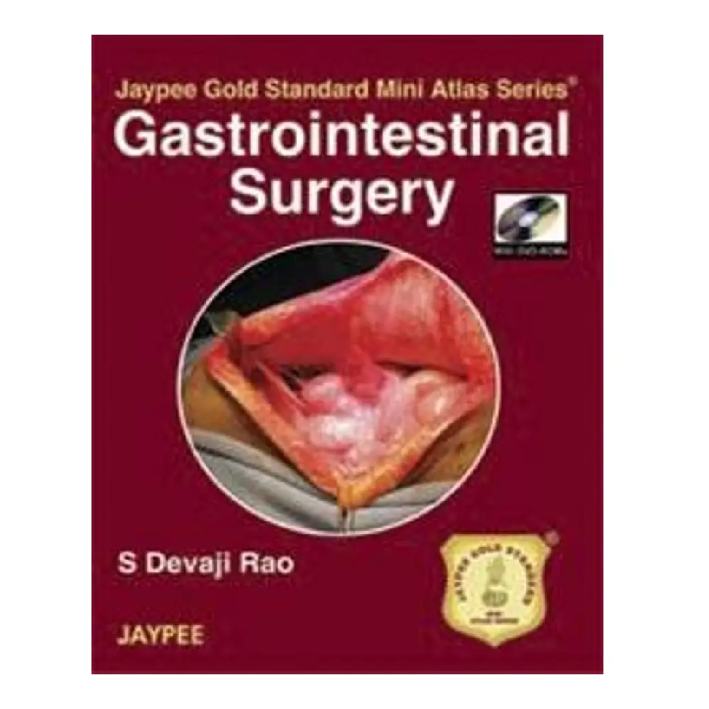
Jaypee Gold Standard Mini Atlas Series Surgical Diseases With Photo CD‑Rom
Jaypee Gold Standard Mini Atlas Series Surgical Diseases With Photo CD‑Rom is a compact visual atlas published as part of the Jaypee Gold Standard Mini Atlas medical book series by Jaypee Brothers Medical Publishers. This atlas is designed to provide high-quality clinical photographs of a wide variety of surgical diseases alongside succinct, practical text describing salient clinical features, diagnosis, and surgical relevance. It serves as an essential visual supplement for students preparing for clinical exams, surgical residents learning pattern recognition, and practicing surgeons revisiting key operative or pathological concepts.
The book is published in paperback format with approximately 304 pages, small enough to fit into a lab coat or backpack, making it useful for quick reference in clinical settings or during revision sessions. A Photo CD-ROM accompanies the printed material, containing additional images that expand the pictorial content and can be used for teaching, presentations, or self-assessment.
Content and Structure
The atlas is organised thematically by surgical specialties and anatomical regions, providing systematic coverage of a broad range of pathological conditions. Rather than lengthy text chapters, the emphasis is on well-labelled, high-resolution clinical photographs showing disease presentations, operative findings, and postoperative results. Key areas include:
1. General Surgery
The atlas begins with core general surgical conditions, showcasing images of common diseases such as appendicitis, hernias, gallbladder pathology, and soft-tissue infections. These images help bridge the gap between textbook definitions and real-world clinical appearances, an especially valuable tool for students who may not have extensive exposure to diverse cases in ward rotations.
2. Head, Neck, and Salivary Glands
Photographs detail surgical presentations involving the neck and salivary glands, such as parotid swellings, submandibular gland stones, and neck masses. Alongside each image, brief summaries highlight clinical signs, differential diagnoses, and key decision points relevant to surgeons.
3. Endocrine and Abdominal Organs
This section covers endocrine surgical disorders like thyroid and parathyroid lesions, as well as intra-abdominal pathology of the pancreas, spleen, adrenals, and digestive tract. The pictures document tumor morphology, inflammation, and procedural steps for common surgical interventions.
4. Breast, Rectum, and Anal Canal
Clinical images of breast lumps, abscesses, rectal lesions, and anal canal conditions help learners recognise both benign and malignant presentations. Particularly for breast diseases, visual correlation with physical findings supports accurate clinical assessments and operative planning.
5. Thoracic and Neurosurgery
Though more specialised, the atlas includes select thoracic surgery and neurosurgical pathology images, as well as brief overviews of relevant surgical techniques. This expands the scope beyond the usual general surgery topics and introduces users to visual features of conditions encountered in broader surgical practice.
6. Urology and Other Techniques
Supplementary sections highlight urological disorders and a few other surgical techniques, offering a more holistic view of surgical disease presentations across specialties.
Photo CD-ROM: Value and Use
One of the atlas’s distinctive features is the Photo CD-Rom included with the print edition. This CD contains a digital repository of images that far exceeds the number of photos printed in the book. These images can be accessed on a computer and are useful for:
-
Teaching demonstrations – instructors can project images during lectures or clinical tutorials.
-
Exam preparation – students can browse, compare, and memorise visual presentations.
-
Case study discussions – trainees can share images in group learning sessions.
While the print version gives immediate access to core visual content, the CD-ROM amplifies the atlas’s educational reach by providing a broader library of clinical photos for self-paced revision.
Audience and Clinical Impact
This atlas is principally aimed at:
-
Medical students – especially those in clinical years preparing for surgery rotations or exams.
-
Surgical residents – who benefit from quick visual reinforcement of disease patterns.
-
Interns and nurses – to improve recognition of surgical pathologies seen on wards.
-
Practising surgeons – as a refresher for less commonly encountered conditions.
The concise style emphasising high-yield visual learning makes it appropriate for busy healthcare professionals who need rapid reinforcement rather than deep theoretical study.
Strengths and Limitations
Strengths
-
Visual focus – the central strength lies in high-quality clinical photographs that show real pathology, aiding pattern recognition.
-
Portable size – compact and easy to carry compared with larger surgical textbooks.
-
Supplemental media – the accompanying Photo CD-ROM significantly increases the amount of visual material available for study.
Limitations
-
Limited text depth – concise summaries are helpful for quick reference but do not replace comprehensive surgical textbooks when deep pathophysiological understanding is required.
-
CD accessibility – reliance on a physical CD may be less convenient given current shifts toward online resources and digital access.
Conclusion
In summary, Jaypee Gold Standard Mini Atlas Series Surgical Diseases With Photo CD‑Rom is a focused, visually rich atlas that enhances learning of surgical diseases through clinical photographs and concise descriptions. Its compact size, practical organisation, and supplemental digital media make it a valuable companion for medical students, trainees, and clinicians seeking to strengthen their visual diagnostic skills in surgery.


Reviews
There are no reviews yet