
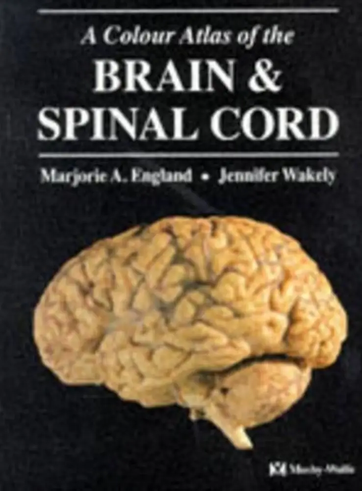

A Colour Atlas of the Brain and Spinal Cord – Overview (600 Words)
A Colour Atlas of the Brain and Spinal Cord is a highly regarded educational resource designed to provide a clear and detailed visual understanding of the human central nervous system. Widely used by medical students, nursing professionals, and healthcare practitioners, this atlas focuses on the structure, organization, and functional anatomy of the brain and spinal cord through high-quality color photographs and concise explanatory text.
The primary strength of this book lies in its visual approach. Unlike traditional textbooks that rely heavily on lengthy descriptions, this atlas uses carefully prepared anatomical specimens and images to help learners grasp complex neuroanatomical concepts quickly. Each image is accompanied by labels and brief notes that explain key structures, making it easier for students to identify and remember important features. This method is especially helpful in a subject like neuroanatomy, which is often considered challenging due to its intricate details and three-dimensional organization.
The book typically begins with an introduction to the basic organization of the nervous system. It outlines the division between the central nervous system (CNS) and peripheral nervous system (PNS), before focusing specifically on the brain and spinal cord. Early chapters may include orientation sections that explain anatomical planes, directions, and terminology, which are essential for understanding subsequent content.
As the atlas progresses, it explores the spinal cord in detail. Cross-sectional images at various levels demonstrate how different tracts and nerve pathways are arranged. These sections help learners understand how sensory and motor information travels between the brain and the rest of the body. The clear labeling of structures such as the gray matter, white matter, and nerve roots allows students to connect theoretical knowledge with actual anatomical visuals.
The brain is covered extensively in later sections. The atlas presents different regions including the cerebrum, cerebellum, and brainstem. Each part is shown from multiple perspectives—external views, internal dissections, and sectional slices. Important structures such as the lobes of the brain, ventricles, cranial nerves, and major pathways are highlighted. By studying these images, learners can develop a spatial understanding of how different parts of the brain relate to each other.
Another valuable aspect of the atlas is its clinical relevance. While the text is concise, it often includes brief clinical notes that link anatomical structures to real-world medical conditions. For example, understanding the location of specific brain regions can help explain symptoms of neurological disorders or injuries. This integration of clinical context makes the book not only informative but also practical for healthcare education.
The atlas is particularly useful for revision and exam preparation. Because of its straightforward layout and emphasis on visuals, it allows students to review large amounts of information quickly. It is often used alongside more detailed textbooks to reinforce learning and improve retention.
In terms of usability, the book is compact and well-organized. The logical progression from simple to complex topics ensures that readers can build their knowledge step by step. The high-quality printing and color images enhance the overall learning experience, making it easier to distinguish between different anatomical structures.
In conclusion, A Colour Atlas of the Brain and Spinal Cord is an essential tool for anyone studying neuroanatomy. Its combination of clear images, concise explanations, and clinical relevance makes it an effective and accessible resource. Whether used for initial learning or quick revision, the atlas provides a solid foundation for understanding the complex structure of the human nervous system.








Reviews
There are no reviews yet